
Whether or not surgery is needed to correct a ruptured cranial cruciate ligament (CCL), rehab is necessary to get an injured dog back to normal again.
Many orthopedic surgeons and rehabilitation veterinarians report that cranial cruciate ligament (CCL) ruptures or tears are the most common limb issues they treat. There are two cruciate ligaments — cranial and caudal — inside the stifle (knee) joint. They connect the femur to the tibia, which is needed for weight bearing. They form an “X” pattern relationship with each other to stabilize the knee.
The cranial ligament is injured the most. This ligament prevents the tibia from gliding forward under the femur, and also prevents internal rotation of the tibia. In dogs, unlike humans, cranial ligament rupture is typically not a result of trauma. The fibers of this ligament may fray over time from wear and tear, and when there is chronic mild inflammation and disease in the joint, enzymes deteriorate and weaken these tissues. Joint instability ensues and leads to osteoarthritis and/or a rupture/tear of the ligament. Genetics and neutering pets too early may cause weak ligaments that can lead to rupture.
Key symptoms to watch for
Typical signs include acute three-legged lameness or toe-touching, and barely any use of the limb. Occasionally, the lameness will be intermittent and chronic, then progressively worsen as the joint disease worsens.
A CCL tear can also lead to injury of the meniscus. This is a cartilage cushion that sits inside the knee and serves as a shock absorber when a dog walks. “If the meniscus tears, it can very painful for the dog and must be treated surgically,” says Kurt Schulz, DVM, MS, DACVS, associate professor of surgical and radiological sciences at the University of California-Davis School of Veterinary Medicine. “About 50% of completely torn cruciate ligaments lead to meniscal injury. When the meniscus is torn, the knee will make a characteristic popping sound when the dog is walking.”
A CCL rupture is usually diagnosed by manipulating the femur and tibia and detecting knee instability. X-rays may determine if there is any pre-existing arthritis and joint effusion, and to look at the positional relationship of the femur and tibia.
Surgical options
Treatment typically involves surgery with post-op rehabilitation. Some patients benefit from rehabilitation and medical management alone, if the condition is not severe, and depending on how unstable the knee is. However, if there is a lot of laxity in the stifle joint and/or a meniscal tear as well, then it is imperative to correct the situation with surgery.
The purpose of any surgery for the cranial cruciate deficient knee is to eliminate too much movement between the femur and the tibia, and remove the whole or part of the meniscus if needed. Because this is a disease and progression usually results in osteoarthritis, correcting the biomechanics of the knee can help slow down degeneration.
There are two categories of such surgeries performed in dogs.
1. Repair with suture/rope material (Lateral Suture Stabilization, Tight Rope). Holes are drilled through the bone to thread synthetic material from the femur to the tibia.
2. Repair with a cut in the bone (Tibial Tuberosity Advancement, Tibial Plateau Leveling Osteotomy) or repositioning the fibula bone (Fibular Head Transposition). TTA and TPLO involve cutting part of the tibia and repositioning it with plates and screws to change the anatomy and biomechanics so the stifle can function without the CCL. FHT surgery repositions the fibula and the attached lateral collateral ligament to mimic the CCL.
Regardless of the type of surgery chosen, arthroscopy to approach the joint should definitely be considered since it is less invasive, and magnification with the instruments helps visualize the anatomy better.
Each patient should be individually evaluated on the severity and chronicity of the situation, and the expected outcome. Then a decision can be made whether to take a surgical approach with post-op rehabilitation protocol, or perform veterinary rehabilitation and medical management only.
The role of rehabilitation
With or without surgery, rehabilitation will benefit the pet by returning the limb to function, hasten recovery from surgery or injury, and improve the patient’s overall body condition, preventing the other CCL from rupturing.
Rehab modalities encompass the following:
• Mechanical — low level laser, therapeutic ultrasound, electric stimulation, shock wave therapy, pulsed electromagnetic fields
• Physical — therapeutic exercises, hydrotherapy, chiropractic, massage
• Energetic — acupuncture, medicinal compounds A combination of these modalities will help decrease pain, inflammation, spasms and edema, and will stimulate repair, improve muscle and nerve tone. Each customized rehab program is done in stages and must advance as the tissues heal and become stronger.
• The focus for the first two weeks after surgical repair or acute injury is to decrease inflammation, pain and swelling. This can be done with a combination of any of the following: cold compression, low level laser, acupuncture, electric stimulation, pulsed therapeutic ultrasound, pulsed electromagnetic field, massage, anti-inflammatories and restricted activity.
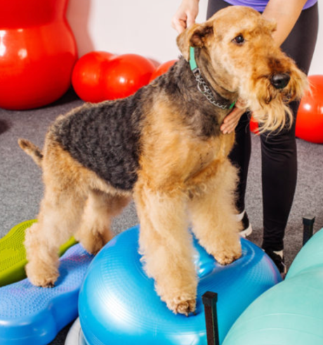
After surgery, depending on the procedure performed, the patient is encouraged to weight shift to restore proprioception, ward off muscle atrophy in the operated leg, and maintain normal posture and range of motion. Once most of the inflammation has subsided, hydrotherapy (underwater treadmill and pool swimming) and easy therapeutic exercises can be introduced.
• By the third week post-op, you want to encourage and increase range of motion, improve limb usage and gait, resolve lameness, halt muscle atrophy, and increase strength, endurance and proprioception. As tissue remodeling occurs and scar tissue is laid down by the body, therapeutic exercises can be made more challenging, with longer, more intense sessions. However, there should be no jumping for about two to four months to allow enough scar tissue to become organized and help stabilize the knee. Chiropractic adjustments are beneficial for correcting subluxations resulting from compensation and long term lameness.
• At about 12 to 16 weeks post-op or post injury, there should be an increase in muscle mass and strength as well as stamina and endurance, an improved use of the limb at higher speeds, and a near normal gait and return to full function (pre-injury level).
Expected outcomes
1. Stifle is free of inflammation.
2. Full range of motion is restored.
3. Limb circumference is symmetrical.
4. Progression of osteoarthritis is delayed.
5. Gait is normal at a walk and trot.
6. Surgery limb is completely healed with improved function, and the opposite limb has improved fitness.
Lifetime management with joint protective nutraceuticals, Omega 3 fatty acids, and natural anti-inflammatories is an important consideration.
As with any physical rehabilitation program, all care should be directly supervised by veterinarians experienced in animal rehabilitation, and performed by a staff of trained animal rehabilitation therapists. In my professional opinion (based on my clinical experience), every pet should have a confirmed diagnosis by a sports medicine orthopedic surgeon or rehabilitation veterinarian before they undergo surgery. The surgical procedure should be performed by a Board Certified Surgeon because his/her level of training and skill set makes him/her the expert in this field.
Author: Julie Mayer, DVM, CVA, CVC, CCRP
Link: https://ivcjournal.com/rehab-for-a-torn-ccl/