

Applying rehab and physiotherapy techniques to a dog with a soft tissue injury should be preceded by a proper consideration of how the injury developed and progressed, and how it affects the health and function of the entire body.
The care of the canine athlete in performance and working disciplines is finally getting much-needed attention and awareness. With the recent growth of agility, flyball, obedience, herding, sled racing and nose-work as competitive canine “hobbies”, more dogs than ever are considered to “have jobs”. Additionally, dogs are working in patrol/protection, detection, search and rescue (SAR), avalanche rescue/recovery and more. Part One of this article (Fall 2018) covered the integrative approach to diagnosing and managing the many musculoskeletal issues that can affect these working dogs, along with specifics about the shoulder. Part Two continues with a focus on injuries involving other body parts.
Carpus and toes
The palmar accessory carpal bone is an important structure in the creation of the palmar carpal canal by acting as a meeting point for several important soft tissues, such as the flexor carpi ulnaris muscle, the palmar carpal metacarpal ligaments, and the flexor retinaculum. The extensor carpi radialis extends across the cranial aspect of the carpus to attach on the proximal metacarpals.
Hyperextension injuries to the carpus are common in active dogs. Most carpal and phalangeal problems are subclinical. Left undetected, they can lead to compensatory problems more proximally in the leg and caudal cervical spine; these are often missed until they are very progressed. Compensation from muscle soreness and tightness in the elbow or shoulder can, in turn, lead to chronic carpal problems. Dogs that are working in deep footing (sand), or that must turn a lot, can develop painful and tight extensor carpi radialis muscles, which can lead to loss of articular congruency and fixations of the carpal bones.

Laxity of the accessory carpal bone can gradually develop from repetitive hyperextension trauma to the supportive soft tissue structures of the palmar carpus; this can occur in dogs that run a great deal (e.g. sled dogs or companion pets that run with the owners frequently). The flexor carpi ulnaris has two heads. The author has had two cases of one head tearing, leading to slight ventral sinking of the carpal joint. Accessory carpal bone laxity can be tested by flexing the carpus approximately 90°, then moving the accessory carpal bone gently back and forth medial to lateral (Image 1). There should be no crepitus, and more movement medially than laterally. The end feel laterally should be very distinctive and firm. In carpal extension, the accessory carpal bone should have a very limited range of motion.
Thermal imaging is an excellent way to initially identify carpal and digital issues. Despite the small size and angularity of most dogs, this area also can be ultrasounded with a small, linear, high-frequency transducer and a stand-off (Image 2).

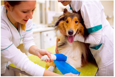
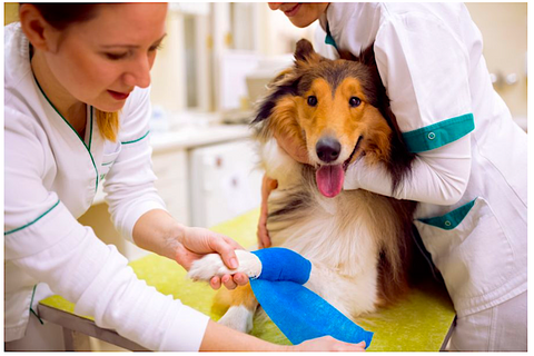
Last but not least, toes should always be examined in the forelimb – always! Toes are usually overlooked, but they’re of critical importance as the interface between the dog and the ground. The foot dissipates load upon landing, and generates propulsion on takeoff. Nail length should be evaluated to make sure they only touch the ground when the foot is active and not when standing still — the practitioner should be able to slide a credit card between the ground and the nail when the dog is standing on a firm, level surface. The length of the toes should also be examined from above; a single digit sticking out longer could be an indication of a torn deep digital flexor tendon. And, of course, the pad surfaces should be carefully examined for any abnormalities or injury (Image 3).

Thermal imaging is an excellent way to identify toe problems; not only can the dorsum of the foot be scanned, but so can the palmar surface. It’s often easier to thermal scan a smooth-surfaced floor where the dog has been standing, than to try and scan the bottom of the foot directly (Image 4). Any dog displaying soreness or heat on thermal imaging should have radiographs taken. Sesamoiditis, phalangeal luxations, phalangeal fractures and flexor tendon damage/ruptures are all common problems in the feet of active dogs. Sesamoiditis is a refractory problem often resolved conventionally by surgically removing the sesamoids. The author has had excellent success using chiropractic to resolve chronic toe and sesamoid injuries.

Lumbosacral spine
For the most part, the working dog’s back (except in the case of iliopsoas issues) rarely shows signs of acute problems except for the odd pulled muscle. Working and performance dogs rarely show disk problems, although spondylosis is common in these dogs as they age. A practitioner is much more likely to see repetitive stress injuries that are gradual and insidious, finally resulting in facet changes and spondylosis. A common diagnosis is “lumbosacral syndrome”. While this term is meant to refer to lumbosacral stenosis, it is often currently used as a “garbage can” diagnosis for generalized back pain.
Integrative practitioners combining the unique palpation skills developed through manipulation and acupuncture examinations, with the advanced imaging of thermography, ultrasound and MRI, are uniquely positioned to fine-tune back pain diagnoses in active dogs. The author has found that many cases labeled “lumbosacral syndrome” ended up being muscles tears, fascial strains and vertebral fixations, with very few actually showing stenosis on MRIs. The majority of these dogs respond well to acupuncture, Chinese and Western herbal use, CBD, manual and manipulative therapies, and core muscle conditioning.
As stated before, many working breeds have such high drive and pain tolerance that they will ignore minor twinges, allowing problems to build until finally a major event occurs. Common guarding signs of back soreness include lumbar kyphosis and loss of “square” neutral stance (the “stable table”). In the dog, neutral stance is evaluated when the front carpi are placed under the elbow and the hind leg metatarsals are perfectly perpendicular to the ground. Any time a dog is seen in a stance or posture other than the “stable table”, with any lumbar kyphosis, he should be evaluated more thoroughly, even if he’s symptom-free. The presence of kyphosis can often be a sign of inadequate core conditioning and strength in a particular dog. Appropriate and flexible core strength is indicated by the dog’s ability to naturally stand in a neutral posture with a neutral (flat) spine. Very athletic and high-drive animals can cheat through their sports with speed and strength rather than using their bodies correctly. If not monitored with proper interventional chiropractic care, improper use will catch up with these dogs, either through aging or when a great effort must be made to avoid or recover from a slip or fall, an event in which tissue strength may be exceeded, leading to injury.
Lumbar lordosis occurs when there is poor core strength. It is often compounded by poor conformation, such as poor hind limb angulation or underlying joint defects like dysplasia. Dogs with lordosis can also benefit from regular manipulative or manual therapy care, combined with appropriate rehabilitation and core conditioning guidance to strengthen the hypaxial and abdominal muscles that counter gravitational sag.
Iliopsoas
Strains and tears of the iliopsoas muscle are more recognized now in the canine athlete; they account for up to 30% of injuries in human sports medicine practices, and are commonly categorized as a groin pull. The presenting signs can be varied, ranging from a dog starting to knock rails while jumping, to chronic vague back pain, to acute weight-bearing lameness. “Iliopsoas” is the name given to the insertional combination of the psoas major and the iliacus muscles on the lesser trochanter of the femur (proximal, medial aspect). In the abdomen, both the psoas major and iliacus are separate muscles. The psoas major originates on various mid-caudal lumbar vertebrae, and the iliacus originates on the cranioventral aspect of the ilium. Both muscles are innervated by the femoral nerve, which originates from the cranial parts of the lumbosacral plexus (L3-L5) and travels through the fused iliopsoas, often making injury to this area painful.

Since the iliopsoas muscle is a hip flexor, an initial diagnosis is made through the clinical test of extending the hind leg behind the dog, then externally rolling the leg out so the stifle swings laterally (and the hip rolls internally). Make sure when the leg is extended that the stifle joint is fully supported by the tester’s hand. A positive response is displayed by an attempt to either pull the leg back or to try to bite the examiner. Additional diagnostics include the use of diagnostic ultrasound by which core lesions and chronic fibrosis are often found in performance canines. Avulsion fractures of the insertion on the lesser trochanter are not common but can be seen on radiographs (Image 5). Chronic core lesions are sometimes treated with ultrasound-guided PRP injections. Avulsion fractures are not surgically repaired.
Regular and routine focused core conditioning, combined with manipulative or manual therapies of the lumbar and sacroiliac areas of the spinal, are critical to keeping the iliopsoas muscle healthy and active and minimizing the potential for injury (as well as caring for the entire lumbar spine). Once an iliopsoas is determined to be injured, it is helpful to be able to stage it with ultrasonography before performing any manipulative therapy. High force chiropractic adjustments should not be applied over any area where there is significant muscular injury (even if suspected) or in the presence of any avulsion or periosteal injury.
Common calcaneal tendon
While full-blown Achilles (common calcaneal) tendon ruptures are uncommon, the increased use of ultrasonography as a musculoskeletal diagnostic tool will probably be able to identify Grade 1 and 2 levels of injury, making earlier recognition more prevalent. In the dog, the calcaneal tendon is made up of three components — the gastrocnemius tendon (the largest component); the common tendons (convergence) of the biceps femoris, gracilis and semitendonosus muscles; and the superficial digital flexor tendon (SDFT). The common tendons make up relatively small insertions of the total calcaneal tendon; the SDFT originates between the two heads of the gastrocnemius muscle and mostly travels on the craniomedial aspect of the gastrocnemius tendon until just proximal to the calcaneus. There, the SDFT fans out and becomes superficial to the gastrocnemius tendon as the SDFT passes over the plantar surface of the tarsal joint. There is also a bursa under the common calcaneal tendon, just proximal to the calcaneus tarsal bone, which can be injured. While most calcaneal tendon injuries are due to acute tendon trauma, associations exist with Cushing’s, obesity, diabetes, and the use of fluoroquinilone antibiotics such as orbafloxacin, enrofloxacin, ciprofloxacin or marbofloxacin. The use of these antibiotics in working and performance dogs is discouraged, although if they’re needed for a life-critical issue, the owners need to be warned that their dogs should not be worked heavily for at least 12 weeks. The author also puts those dogs on vitamin C protocols for at least three to six months to support tropocollagen production. Common calcaneal tendon (CCT) insertion strain in the opposite weight-bearing limb can commonly result from a non-weight-bearing lameness.
Significant proprioceptive nerve fibers serve the calcaneal tendon. Research in humans has shown that vibration of the calcaneal tendon has a major impact on stance and posture. This is due to the stimulation of both muscle spindle cells (MSCs) and Golgi tendon organs (GTOs) in the numerous muscles that combine to make the calcaneal tendon. In humans, mechanical stretching of the common calcaneal tendon results in a stance that leans posteriorly in anticipation of forward movement. The calcaneal tendon may be just as, or even more important, in a quadruped. Body work and structural rebalancing of any tissue involved with the hindlimb is sure to make profound rebalancing changes to the hindquarters (and no doubt also to systemic movement).
Calcaneal tendon injuries other than ruptures typically have a vague history of low grade, consistent or off-and-on lameness. Rarely is there palpable thickening, heat or sensitivity at the area of the lesion. Diagnosis is made by identifying the location using thermal imaging, then documenting it with ultrasonography. Chronic-active injuries should be approached with caution when applying any manipulative force across the area, due to the possibility of fiber degeneration at the periosteal insertion. If the tissue shows itself able to handle manual or acupuncture modalities, these can positively affect the hind limb neuromuscular and neurofascial tissue dialogue.

Tarsus/metatarsals
The tarsus in the athletic dog is usually overlooked. Often, abnormal radiographic appearance is not recognized (Image 6). Like the carpus, the tarsus is a complex joint that dissipates shearing and concussive stresses by transferring them through the large cartilaginous surface area created by the small bones. There are numerous small ligaments, as well as the larger plantar fascia/ligament on the caudal aspect of the distal tarsus, the flexor tendons and the interosseous muscle (equivalent to human plantar fascia and equine suspensory ligament). Recognizing potential injuries in this area is important. Racing greyhound sports medicine recognizes and has published papers on several conditions of the tarsus, many of which are also seen in active companion and working dogs. Recently, the author treated a racing dog at the Iditarod for a lateral collateral tarsal ligament injury; early recognition and prompt stabilization resulted in a rapid recovery and return to full function.
 The plantar interosseous muscle and individual digital flexor muscles can also be injured. These can be recognized through very soft and thorough palpation (create a picture with your fingers), and/or thermography and diagnostic ultrasonography (Image 7).
Stifle (femorotibial joint) ligament injuries
Stifle joint ligament injuries include those of the cranial cruciate, medial and lateral collateral ligaments. Not every case of hind limb lameness involves the cruciate ligament. Groin muscles (especially of the adductor insertional tendon) and collateral ligaments can also look like stifle injuries. When testing the stifle, make sure that both the cranial drawer and the tibial thrust tests are performed, with the dog both standing and lying down as well as with the limb extended and flexed – this can help determine the status of the cranial cruciate ligament (partial or full tear).

It is beyond the scope of this article to discuss surgical repairs to the canine CCL, but consider surgical options mindfully. Current surgical techniques seem to be suggested almost too casually, and are not without long-term problems. The cruciate ligament
probably plays a significant role in total limb proprioception and “joint position sense”. Ligamentous injury that leads to laxity in the human knee joint (animal stifle joint) has been shown to not only affect the injured leg, but can also set the body up for kinematic differences in other joints (including in the unaffected leg) as well as for systemic proprioceptive loss and significant gait asymmetries. Several human studies have reported a loss of proprioceptive function and increased laxity in the normal uninjured knee in
conjunction with an ACL injury.
Unlike in the active human, not much research has been done in dogs to examine the potential involvement of complex biomechanical forces and events being involved in stifle injuries. Dogs can definitely injure the propulsion (hamstring complex) and braking (quadriceps, tensor fascia lata) muscles of the hind leg. More thorough examination recording, combined with the use of soft tissue imaging such as thermography and diagnostic ultrasound, may reveal what many integrative sports medicine practitioners report – many active dogs that eventually have a cranial cruciate ligament failure have also had previous chronic muscular injuries. How could one not affect the other? Integrative practitioners, especially those trained in chiropractic, osteopathy and myofascial techniques, could be on the front line of developing better early-warning protocols for stifle ligament injuries.
Integrative treatment approaches
It seems to be the nature of today’s medicine to rely on recipe-like checklists for treatment protocols. The introduction of rehabilitation seems to very much rely on predictable and consistent protocols for post-injury treatment. The success of such protocols can easily lead practitioners to ignore or miss individual variations and responses to treatment.
Integrative practitioners have numerous tools to meet the individual needs of a patient. Acute injury tools such as cold laser, acupuncture, homeopathy and herbs can be very effective in all phases of healing. Structural alignment, manual and manipulative therapies can be easily integrated into rehabilitation visits and protocols, adding supportive therapies to healing that assist in speeding up recovery by removing tissue stressors from imbalanced movement, blood flow restrictions and neurologic impingement. There unfortunately isn’t the space in this article to go through each and every modality available to those working with the management of canine athletes. But, as with any veterinary approach, it is just as imperative with integrative therapies as with conventional ones to first make an accurate and through diagnosis. Practitioners are encouraged to review their modalities for techniques specific to muscular, fascial, ligamentous, tendinous, neurologic and skeletal injuries.
Conclusion
Injuries sustained during activity can be as complex and varied as life itself; integrative therapies recognize the picture of the individual more than conventional, reductionist modalities do. The combination of all perspectives, conventional and integrative alike, provides the practitioner with a larger picture of the individual within the population. As with any veterinary treatment, it is just as imperative with integrative therapies to first have a diagnosis. The goal of this article has been to encourage rehabilitation, integrative, and even conventional practitioners to take a more focused and detailed diagnostic look at any animal that presents with lameness, no matter how vague. All practitioners should be willing to create a bigger and better picture for any individual patient through a more detailed and knowledgeable approach.
Author: Kimberly Henneman, DVM, DACVSMR (EQ, K9), FAAVA, DABT, CVA, CVC
Link: https://ivcjournal.com/recognizing-soft-tissue-injuries-dog-2/