

Radiology is one of the most widely used and accessible imaging techniques for evaluating the musculoskeletal system in dogs, but interpreting and translating these findings into effective treatment plans can sometimes be challenging. In this lecture and lab, we will cover the basics of musculoskeletal radiographic interpretation, focusing on commonly assessed joints such as the hip, stifle, elbow, and shoulder. We will emphasize clinically relevant tips for interpreting radiographs, alongside recent literature to guide our readings. Additionally, other imaging modalities like ultrasound, CT, and MRI may be indicated based on initial findings, and we will discuss appropriate next steps. The core focus will be on translating radiographic results into actionable rehabilitation and modality management strategies. Through case-based discussions and hands-on exercises, students will learn to develop tailored rehab plans that address the specific needs of each patient, incorporating various therapeutic modalities to optimize recovery and function. Live canine patients with radiographs will be presented, allowing students to collaboratively review the images and apply appropriate treatments based on both the radiographic findings and the patient’s clinical presentation.
Lecture Overview: 1 hour and 15 minutes
Brief review of general radiographic interpretation basics and changes associated with musculoskeletal disease. Cover key radiographic landmarks, signs of pathology, and evidence-based interpretation strategies. The core of this course centers on translating imaging findings into effective rehabilitation plans. Current literature will be incorporated to support clinical decision-making, and we’ll discuss when additional imaging is indicated to further clarify diagnoses or guide treatment. The focus will be on the hip, stifle, shoulder, and elbow. Topics will include:- Correlation radiographic abnormalities with clinical signs and physical exam findings.
- Choosing appropriate therapeutic modalities based on imaging.
- Designing rehabilitation plans tailored to the patient’s specific condition and functional goals.
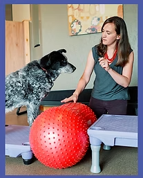
Hands-On Lab: 2 hours and 45 minutes
4 cases (40 minutes per case) - ideally shoulder pathology case, elbow pathology case, hip pathology case, and stifle pathology case. Each patient will have a history and brief physical exam, radiographs to review as a group collaboratively and develop a treatment plan.- Practice reading musculoskeletal radiographs in a clinical context.3
- Collaboratively assess patient presentation alongside imaging findings.
- Develop and apply individualized rehabilitation plans using appropriate modalities.


Reviews
There are no reviews yet.