

Seven measurement tools and methods veterinarians can implement into equine rehabilitation programs to track progress.
As we know, the list of rehabilitation modalities currently used in horses is far longer than the row of scientific articles lined up to support their use.
“While it is widely accepted that equine rehabilitation not only helps prevent injury but also restores function following an injury and enhances a horse’s athletic performance, the practice of evidence-based medicine mandates valid outcome measures,” Connor said. “Otherwise, we are unable to provide credible and reliable justification for these therapies and are unable to devise specific therapeutic protocols for particular indications.”
In other words, without the science backing up these programs, the industry remains reliant on testimonials touting the virtues of rehabilitation … much like what owners encounter when attempting to purchase oral nutritional supplements for their horses.
“Having objective baseline functional values, such as the maximum flexion or extension of a joint, circumference of an effusive (swollen) joint, or forelimb retraction length, for each patient allows the therapy protocol to be altered during treatment to ensure the desired effect on the target tissue is achieved,” said Connor.
Heat, pain, and swelling are hallmark features of most equine injuries, and they can contribute to decreased range of motion. Many of the available tools to objectively measure the extent of an injury measure these parameters. Connor provided the following examples of outcome measures veterinarians can implement into equine rehabilitation programs:
1. Goniometry This method uses a protractorlike tool with extendible arms to measure a joint’s range of motion (flexion, extension). The veterinarian must place the goniometer at the joint’s center of rotation.
“Goniometry measures progress in joint mobility and can be easily integrated into rehabilitation protocols that use exercises like stretching and passive joint mobilization to enhance soft tissue extensibility,” Connor explained.
2. Pressure algometry Due to the development of radiating (starting in one area and spreading to a larger one) and referred (perceived at a location other than the site of the painful stimulus) pain, localizing the primary source of musculoskeletal pain can be challenging in equine medicine. Pressure algometry helps measure “mechanical nociceptive thresholds” (MNTs) to quantify pain and track whether it worsens or improves. This handheld unit applies localized pressure to a specific part of the horse’s body. The lower the amount of pressure needed to cause the horse to withdraw, the more pain the horse feels.
“As a horse progresses through their rehabilitation program, the MNTs should increase, demonstrating that more pressure is required to elicit a painful response,” said Connor.
3. Thermography As mentioned, heat commonly occurs with injuries as a natural part of the inflammatory process. Increased blood flow to an injury can increase skin temperature, which veterinarians can easily measure using a small thermography unit. To provide useful data, the practitioner must use thermography under similar conditions each time (e.g., ambient temperature, same location on the horse’s body, after the same amount of work/exercise has been performed) and should always compare results to the contralateral (opposing) body part.
4. Malleable ruler Rather than eyeballing symmetry over the back and hindquarters, this flexible ruler contours the horse’s body and can be used to create a tracing of the horse’s body onto graph paper. Veterinarians can measure these tracings to identify asymmetry and abnormalities and track measurements moving forward to assess progress, such as muscular improvement.
5. Lateral bending Veterinarians can use several methods, such as photographs or direct measurements with a tape measure, to measure the distance between a horse’s nose and the desired landmark, such as the hip, hock, and girth.
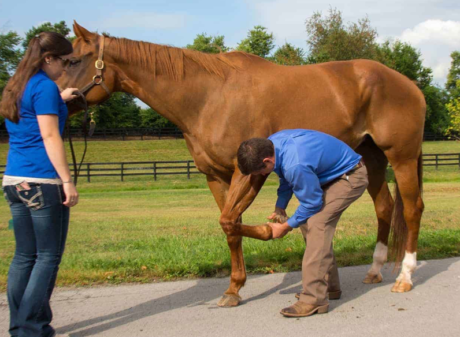
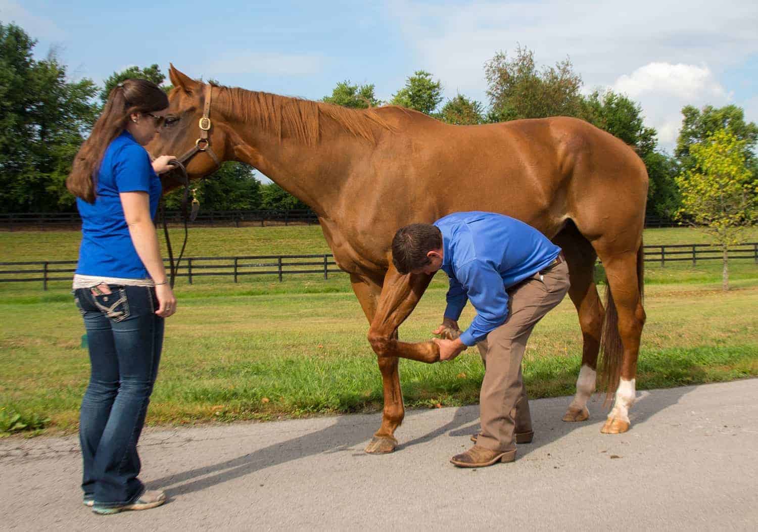
6. Retraction length Simply measuring the length of a straight line on the floor from one forelimb in a square position to that same limb maximally retracted can provide valuable information regarding forelimb muscle responses to therapy.
7. Circumferential limb or joint circumference These data provide information on swelling (e.g., joint effusion will increase joint circumference and should decrease over time with rehabilitation therapy) and muscle mass.
Author: Stacey Oke, DVM, MSc
Link: https://thehorse.com/186141/how-to-measure-horses-rehabilitation-progress-objectively/